

The Patient
Scottie, a 6 year old male neutered Boxer, came to see me after his owner found a small lump on his skin.
The Case
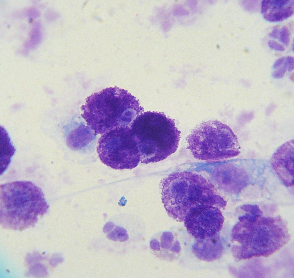
On examination, I found a roughly 1cm round slightly raised hairless dermal (skin) mass located on the inside surface of Scottie's right thigh. It did not seem to bother him but there was hair loss over the mass and it was slightly pink in color. I also found a second mass located on the outside surface of the same leg near the ankle. It had a very similar appearance but was a little smaller. I recommended that I take a fine needle aspirate (FNA) of the two masses for cytology to make a diagnosis. This entails using a small needle to try to get some cells from the mass to look at under the microscope. Scottie was a very good boy and held very still (with the help of some yummy cheese) while I took the aspirates. I then made a slide of the sample and looked at it under the microscope. Each mass looked exactly the same. The slide was covered in uniform round cells with darkly pigmented granules inside the cells' cytoplasm. These types of cells are called mast cells and when they are found in a large quantity inside a lump, this is called a mast cell tumor (MCT). Mast cells are part of the immune system and release histamine as part of an allergic reaction (think of a hive that develops on the skin after a bee sting). Unfortunately, mast cell tumors are always malignant, meaning they have the potential to metastasize (spread) to lymph nodes and other organs such as the liver or spleen. There are different grades of MCTs indicating their propensity to spread throughout the body but we can't know the grade based just on cytology. This requires a biopsy.
The Treatment Plan
I recommended that we schedule Scottie to have surgery to remove the two masses and submit them for biopsy. Whenever I am removing a known MCT, I always try to take at least 2-3cm of normal skin surrounding the mass to make sure we get the whole mass so it doesn't just grow back. This means a larger incision than most owners expect. Thankfully, incisions heal from side-to-side not end-to-end so a 1cm incision heals just as fast as a 3cm incision. The owner approved and we scheduled the surgery for the following week. We also collected a blood sample to make sure he was healthy enough for anesthesia and surgery. I also talked to her about post-op care and pain management following the surgery. The following week, Scottie arrived for his surgery. He was placed under general anesthesia and given appropriate analgesia (pain control) both pre-op and intra-op. I made an elliptical shaped incision around each mass with 2-3cm margins and then closed the incisions with 2 layers of sutures (stitches). The lumps were placed in formalin and submitted to the lab for histopathology (biopsy). Scottie stayed in the hospital all day for monitoring and pain management and sent home around 5pm. I met with his owner to go over all of my post-op instructions and his medications. He needed to wear an Elizabethan collar (E-collar/cone) until the sutures were to be removed in 10-14 days. This is super important as dogs are really good at chewing out their sutures before it is time for that.

The Outcome
Scottie did very well and made a full recovery. I received the biopsy results back in about 7 days. The two masses were grade 2 MCTs with clean margins. This means that we got the whole tumor and they have a low risk for metastasis. No further work-up or treatment was indicated. I did let the owner know that Scottie could get another MCT in the future so to bring him in immediately if she found a new mass. Sadly, Boxers have a higher risk for this type of tumor than most breeds so I see this very commonly for them. The sooner a MCT is removed, the better the prognosis. MCTs can be very sneaky as they do not always look the same on examination. Sometimes they are on the skin (like Scottie) but sometimes they are under the skin, called subcutaneous, and feel like a fatty or cystic mass. Sometimes they are pink and hairless (like Scottie) but sometimes they are darkly pigmented and smooth. They are "the great pretenders" of skin tumors. This highlights the importance of sampling all masses to ensure an accurate diagnosis. If you find a mass on your dog's body, be sure to schedule an appointment with us soon.
The Drake Center for Veterinary Care is an AAHA-accredited animal hospital located in Encinitas, CA. The Drake Center loves being a source of information for all pet owners across the country; however, if you have any questions regarding pet care and do not live in Encinitas, CA or surrounding cities, we encourage you to contact your local veterinarian.